Types of Skeletons in Living Things
Theme: THE HUMAN BODY
Topic 1: THE MUSCULAR AND SKELETAL SYSTEM
Subtopic 1: Types of Skeletons in Living Things • Source: Primary 7 Science National Curriculum Textbook & P.7 Science Comprehensive Guide
1. Introduction to Skeletons
In all living organisms that possess body form, support is required to maintain shape, protect soft tissues, and enable purposeful movement.
A skeleton is the supporting structure of the body of an organism.
❌ Mistake: Defining a skeleton as "a framework of bones in the body".
✔ Examiner Correction: "A framework of bones in the body" is specifically the human skeleton or vertebrate endoskeleton. Insects and worms have skeletons but possess no bones at all. In Section A examinations, always write: "The supporting structure of the body of an organism."
2. The Three Types of Skeletons in Living Animals
Animals in the animal kingdom possess three distinct types of skeletons based on their structural location and composition:
A. Exoskeleton (External Skeleton)
- Definition: An exoskeleton is a skeleton found on the outside of the body of an animal.
- Composition: It is made of a tough, non-living, waterproof outer protective covering called a cuticle, containing the tough substance chitin.
- Functions of an Exoskeleton:
- Provides structural support and gives shape to the animal's body.
- Protects the delicate soft internal organs from physical injury and predator attacks.
- Prevents excessive water loss (desiccation) from the animal's body, enabling survival in hot, dry environments.
- Provides points of attachment for internal muscles that move the legs and wings.
- Growth Limitation and Moulting (Ecdysis):
Because the exoskeleton is a rigid, non-living structure, it cannot expand or grow continuously. To increase in size, arthropods must periodically shed their old outer cuticle in a biological process called moulting (ecdysis).
During moulting, the animal swallows air or water to expand its body, cracks the old tight cuticle, crawls out, expands rapidly while the new soft skin underneath is flexible, and then allows the new cuticle to harden.
- Examples of Animals with an Exoskeleton (All Arthropods):
- Insects: Houseflies, honeybees, grasshoppers, locusts, cockroaches, butterflies, termites, mosquitoes, and beetles.
- Arachnids: Spiders, scorpions, ticks, and mites.
- Myriapods: Millipedes and centipedes.
- Crustaceans: Crabs, lobsters, woodlice, crayfish, prawns, and shrimps.
B. Endoskeleton (Internal Skeleton)
- Definition: An endoskeleton is a skeleton framework found inside the body of an animal.
- Composition: It is composed of hard, living biological tissues called bones and flexible tissues called cartilage.
- Continuous Growth: Unlike an exoskeleton, an endoskeleton is made of living cells supplied with blood vessels and nerves. It grows continuously and proportionally with the animal's body, so vertebrates do not undergo moulting.
- Functions of an Endoskeleton:
- Supports the body weight against the force of gravity.
- Provides a firm internal framework that gives the animal its characteristic shape.
- Shields and protects delicate internal body organs (brain, spinal cord, heart, lungs).
- Works with voluntary skeletal muscles to produce coordinated movement and locomotion.
- Stores essential minerals (primarily calcium and phosphorus).
- Manufactures red blood cells, white blood cells, and platelets inside the red bone marrow.
- Examples of Animals with an Endoskeleton (All Vertebrates):
- Mammals: Humans, cows, goats, dogs, horses, elephants, whales, and bats.
- Birds: Ostriches, eagles, domestic fowl, ducks, and weavers.
- Reptiles: Lizards, crocodiles, snakes, and tortoises (Note: Tortoises possess an endoskeleton inside, surrounded by a bony shell / carapace).
- Amphibians: Frogs, toads, newts, and salamanders.
- Fish: Tilapia, Nile perch, mudfish, lungfish, and sharks (cartilaginous skeleton).
C. Hydrostatic Skeleton (Fluid-Pressure Skeleton)
- Definition: A hydrostatic skeleton is a skeleton in which an animal's soft body is supported and kept rigid by body fluids held under pressure inside closed body cavities (the coelom).
- Mechanism of Movement: The animal possesses layers of circular and longitudinal muscles in its body wall. When circular muscles contract, the fluid pressure pushes the body outward making it long and thin; when longitudinal muscles contract, the body becomes short and thick. This hydraulic action allows locomotion through soil or water.
- Examples of Animals with a Hydrostatic Skeleton:
- Annelids: Earthworms and leeches.
- Molluscs (soft parts): Snails, slugs, octopuses, and squids.
- Insect Larvae: Caterpillars, maggots, and cutworms.
- Cnidarians & Others: Jellyfish, sea anemones, starfish, and roundworms.
3. Detailed Comparison Matrix of Skeleton Types
| Feature | Exoskeleton | Endoskeleton | Hydrostatic Skeleton |
|---|---|---|---|
| Location | Outside the body | Inside the body | Internal fluid-filled cavity |
| Composition | Non-living chitin / cuticle | Living bones and cartilage | Coelomic fluid under pressure |
| Growth Mode | Intermittent through moulting (ecdysis) | Continuous with body growth | Continuous fluid expansion |
| Animal Group | All Arthropods (Insects, Spiders, Crabs) | All Vertebrates (Mammals, Birds, Fish) | Soft-bodied Invertebrates (Worms, Snails) |
| Major Advantage | Prevents water loss; hard armor | Supports large body mass; grows inside | High flexibility; easy burrowing |
- Write Exoskeleton as ONE word (not "Exo-skeleton" or "Exo skeleton").
- Write Endoskeleton as ONE word (not "Endo skeleton").
- Write Hydrostatic skeleton as two words with correct spelling.
- Write Ecdysis or Moulting when asked for the process of shedding the cuticle.
Structure of the Human Skeleton & Bone Classification
Theme: THE HUMAN BODY
Topic 1: THE MUSCULAR AND SKELETAL SYSTEM
Subtopic 2: Human Skeleton Architecture & Bone Classification • Source: Primary 7 Science National Curriculum Textbook & P.7 Science Comprehensive Guide
1. Architecture of the Human Skeleton
The human skeleton is an endoskeleton constructed of bones, cartilage, and strong connective tissues.
- An adult human skeleton contains exactly 206 distinct bones.
- A newborn baby is born with approximately 300 to 305 bones and cartilages.
- Why do babies have more bones than adults? Babies are born with many separate, pliable cartilaginous pieces. As the child grows, these cartilages absorb calcium and phosphorus, fuse together, and turn into solid bone through a biological process called ossification.
- The longest, strongest, and heaviest bone in the human body is the Femur (thigh bone).
- The smallest bone in the human body is the Stirrup (Stapes) located in the middle ear.
2. Functions of the Human Skeleton
- Protection of Delicate Internal Organs:
- Skull / Cranium: Protects the brain, eyes, tongue, inner ear, and middle ear.
- Vertebral Column (Backbone / Spine): Protects the spinal cord.
- Rib Cage (Ribs & Sternum): Shields and protects the heart and lungs.
- Pelvic Girdle (Pelvis / Hipbone): Shields and supports the urinary bladder and reproductive organs.
- Eye Sockets (Orbits): Protect the eyeballs from physical blows.
- Mandible (Lower Jaw): Protects the tongue and anchors lower teeth.
- Support: Forms a rigid structural scaffold that carries and bears the entire weight of the upper body.
- Body Shape & Framework: Gives the human body its distinctive upright, erect posture and physical form.
- Locomotion and Movement: Acts as a mechanical lever system; skeletal muscles attach to bones and pull on them to move the limbs.
- Manufacture of Blood Cells (Hematopoiesis): Red bone marrow found inside spongy bone and flat bones manufactures red blood cells, white blood cells, and platelets.
- Storage of Essential Minerals: Bones act as an internal storehouse for mineral salts, predominantly calcium and phosphorus, which keep bones strong and rigid.
3. Major Divisions of the Human Skeleton
The 206 bones of the human skeleton are systematically divided into two main divisions:
A. The Axial Skeleton (Central Axis)
The axial skeleton comprises the central longitudinal axis of the body (80 bones):
- The Skull (Cranium & Facial Bones): 22 bones.
All skull bones are fused tightly together by immovable fibrous joints called sutures, EXCEPT the mandible (lower jaw bone), which is the ONLY movable bone in the skull.
- The Vertebral Column (Backbone / Spine):
Consists of 33 irregular bones called vertebrae, arranged into five specific regions:
| Vertebral Region | Number | Location & Special Features |
|---|---|---|
| 1. Cervical Vertebrae | 7 | Neck region. C1 = Atlas (supports skull, allows nodding) and C2 = Axis (allows head rotation/turning). |
| 2. Thoracic Vertebrae | 12 | Chest region. Each connects behind to one of the 12 pairs of ribs. |
| 3. Lumbar Vertebrae | 5 | Lower back / loins. Largest, thickest, and strongest unfused vertebrae; bear body weight. |
| 4. Sacral Vertebrae (Sacrum) | 5 (fused) | Pelvic region. Fused together into a single, strong triangular wedge bone. |
| 5. Caudal / Coccyx | 4 (fused) | Tailbone. Fused together at the base of the spine; remnant of ancestral tail. |
| TOTAL VERTEBRAE | 33 | Formula: 7 + 12 + 5 + 5 + 4 = 33 Bones |
- The Rib Cage:
Consists of 12 pairs of ribs (24 ribs total) and the central breastbone called the Sternum:
- True Ribs (Pairs 1 to 7): 7 pairs connected directly to the sternum in front via their own costal cartilages.
- False Ribs (Pairs 8, 9, 10): 3 pairs attached indirectly to the sternum by fusing into the costal cartilage of the 7th rib.
- Floating Ribs (Pairs 11 and 12): 2 pairs attached only to the thoracic vertebrae behind, but completely free and unattached in front.
B. The Appendicular Skeleton (Limbs & Girdles)
Consists of the 126 bones of the appendages and their anchoring girdles:
- Pectoral Girdle (Shoulder Girdle):
- Clavicle (Collarbone): Slender, curved S-shaped rod holding the shoulder away from the chest.
- Scapula (Shoulder blade): Flat, triangular bone on the upper back that forms the cup socket for the humerus.
- Forelimbs (Upper Limbs / Arms):
- Humerus: Upper arm bone; longest bone in the upper limb.
- Radius: Forearm bone on the lateral (thumb) side.
- Ulna: Forearm bone on the medial (pinky finger) side.
- Carpals: 8 short wrist bones forming a gliding joint.
- Metacarpals: 5 palm bones.
- Phalanges: 14 finger bones (2 in thumb, 3 in each of the four fingers).
- Pelvic Girdle (Hip Girdle / Pelvis):
Composed of three pairs of fused hipbones (ilium, ischium, pubis). Transmits upper body weight to the legs and protects the reproductive organs and bladder.
- Hindlimbs (Lower Limbs / Legs):
- Femur (Thigh bone): Longest and strongest bone in the human body.
- Patella (Kneecap): Small sesamoid bone protecting the front of the knee hinge joint.
- Tibia (Shinbone): Larger, thicker, inner weight-bearing bone of the lower leg.
- Fibula: Thinner, outer bone of the lower leg providing muscle anchoring.
- Tarsals: 7 short ankle bones forming a gliding joint.
- Metatarsals: 5 bones forming the sole / instep of the foot.
- Phalanges: 14 toe bones (2 in big toe, 3 in each of the four other toes).
4. Classification of Bones by Shape
- Long Bones: Elongated bones with a central shaft and swollen articulating ends. Contain yellow marrow in the shaft and red marrow at the ends. Examples: Femur, humerus, tibia, fibula, radius, and ulna.
- Short Bones: Small, cube-shaped bones that contain red bone marrow. Examples: Carpals (wrist) and tarsals (ankle).
- Flat Bones: Thin, broad, flattened bones that protect internal organs. Examples: Cranium (skull), scapula (shoulder blade), sternum (breastbone), pelvis, and ribs.
- Irregular Bones: Complex, non-uniform shapes providing specialized protection and support. Examples: Vertebrae (backbone) and pelvic bones.
5. Detailed Longitudinal Structure of a Long Bone
A typical long bone (such as the femur or humerus) exhibits distinct histological layers and compartments:
- Periosteum:
A tough, fibrous outer membrane covering the entire exterior of the bone (except at joints).
Functions: Protects the bone, provides attachment points for muscles and tendons, and contains blood vessels that supply oxygen and nutrients to bone cells. - Compact Bone:
A very dense, solid, and hard outer layer of bone tissue located beneath the periosteum.
Functions: Gives the bone rigidity, mechanical strength, and enables it to withstand heavy loads and physical impacts. - Spongy (Cancellous) Bone:
A honeycomb-like porous mesh of bone tissue located inside the swollen ends (epiphyses) of long bones.
Functions: Reduces the overall weight of the skeleton and houses the red bone marrow. - Red Bone Marrow:
Specialized blood-forming tissue located within spongy bone cavities, short bones, and flat bones.
Function: Manufactures red blood cells, white blood cells, and blood platelets (hematopoiesis). - Medullary Cavity (Marrow Cavity):
A hollow central cylinder running through the shaft (diaphysis) of long bones.
Function: Filled with yellow bone marrow, which stores fat and lipid energy reserves. - Articular Cartilage:
A layer of smooth, resilient, elastic connective tissue capping the joint ends of long bones.
Functions: Absorbs mechanical shock, cushions bone ends, and prevents bones from rubbing directly against each other, eliminating friction and wear.
Joints in the Human Body & Supporting Structures
Theme: THE HUMAN BODY
Topic 1: THE MUSCULAR AND SKELETAL SYSTEM
Subtopic 3: Articulations, Joint Mechanics & Connective Tissues • Source: Primary 7 Science National Curriculum Textbook & P.7 Science Comprehensive Guide
1. What is a Joint?
A joint is a place in the body where two or more bones meet and articulate.
Primary Functions of Joints: Provide body flexibility, allow smooth movement, and enable coordinated locomotion.
2. Classification of Joints in the Human Body
Joints are categorized into two primary functional classes:
A. Immovable / Fixed / Fibrous Joints
- Characteristics: The articulating bones are interlocked and held tightly together by tough, fibrous connective tissue. They allow no movement whatsoever.
- Primary Example: The suture joints of the cranium / skull. The jagged edges of the skull plates interlock like puzzle pieces to provide maximum rigid protection for the delicate brain.
- Other Example: Sockets of the jaw holding the teeth (gomphoses).
B. Movable / Synovial Joints
Joints that allow free movement between articulating bones. In Primary 7 Science, there are four major types of movable joints:
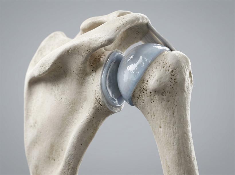
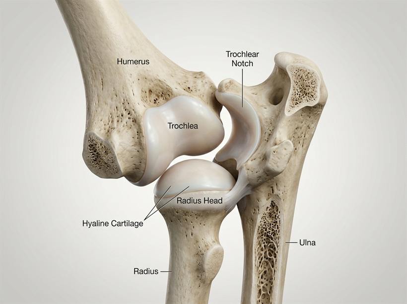
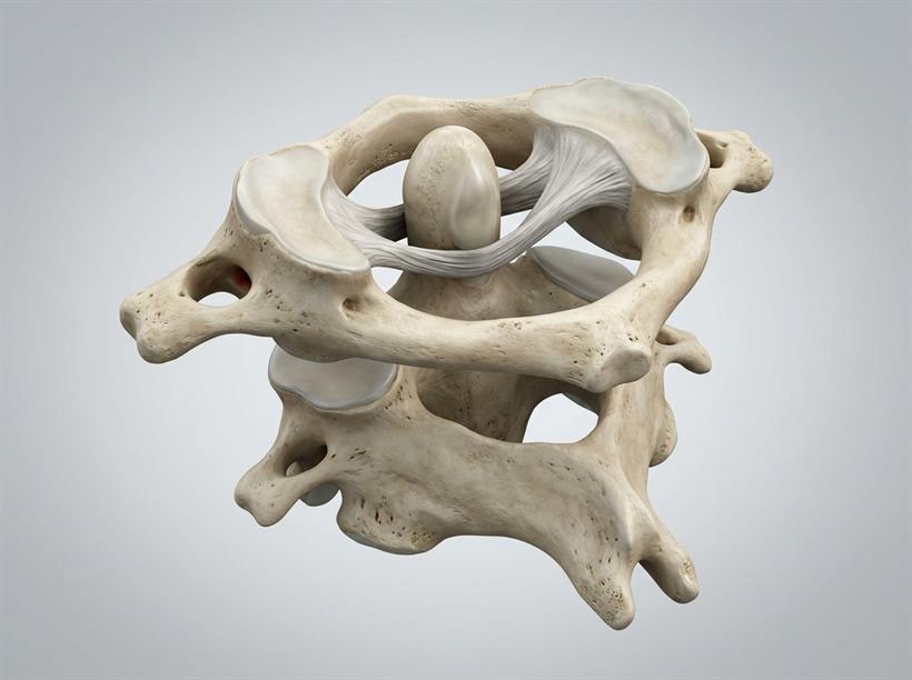
| Type of Joint | Plane of Movement | Locations in the Body | Special Features |
|---|---|---|---|
| 1. Hinge Joint | Movement in only one plane / direction (180°, like a door hinge). | • Elbow joint • Knee joint • Knuckle / finger joints (phalanges) |
Allows bending (flexion) and straightening (extension). |
| 2. Ball and Socket Joint | Movement in all directions / three planes (360° omnidirectional rotation). | • Shoulder joint (humerus in scapula) • Hip joint (femur in pelvis) |
Formed by a rounded ball head fitting into a cup-shaped cavity; offers greatest mobility. |
| 3. Pivot / Rotary Joint | Allows one bone to rotate or pivot on another. | • Neck joint (between Atlas C1 and Axis C2 vertebrae) | Enables the head to nod up/down, rotate side-to-side, and shake. |
| 4. Gliding / Plane Joint | Allows flat bone surfaces to slide smoothly over each other in two planes. | • Wrist joint (between carpals) • Ankle joint (between tarsals) |
Provides fine, flexible hand and foot adjustments during walking and writing. |
3. Detailed Anatomy of a Movable (Synovial) Joint
To withstand constant movement and heavy mechanical pressure, movable joints contain specialized protective tissues:
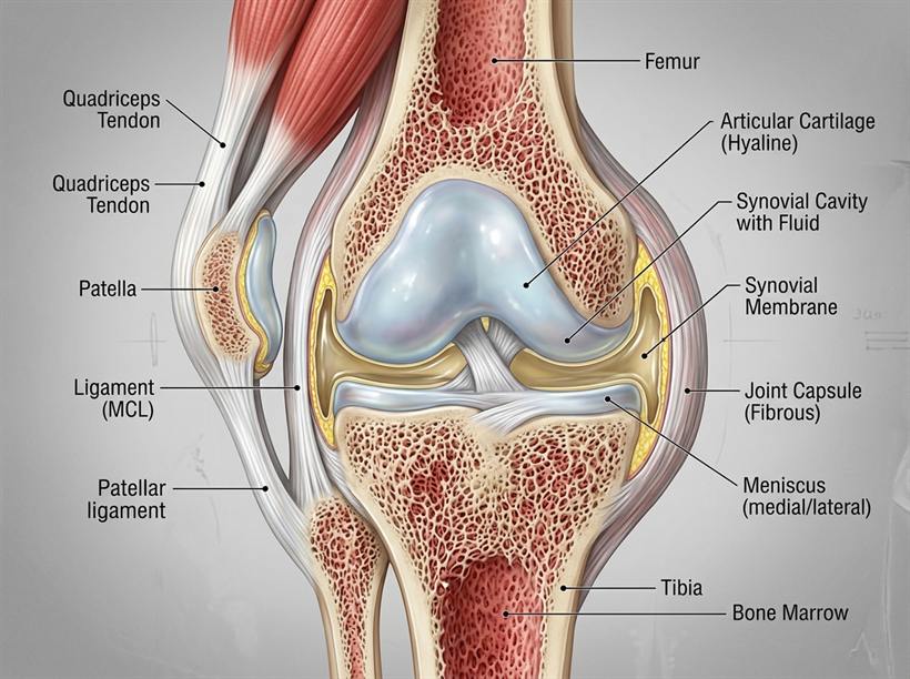
- Ligament:
A band of tough, elastic fibrous connective tissue.
Function: Joins / attaches bone to bone at a joint, stabilizes the joint, and prevents the bones from dislocating during vigorous movement. - Tendon (Sinew):
A band of tough, inelastic dense white fibrous collagen tissue.
Function: Attaches muscle to bone and transmits the mechanical pull of muscle contraction to move the bone. - Cartilage (Articular Cartilage / Gristle):
A smooth, soft, tough, and rubbery connective tissue capping the articulating ends of bones.
Functions: Cushions bone ends, absorbs mechanical shocks during jumping or walking, and prevents bones from rubbing directly against each other, eliminating friction and wear. - Synovial Membrane:
A thin lining layer of tissue that encloses the joint capsule.
Function: Secretes, replenishes, and holds the synovial fluid. - Synovial Fluid:
A clear, slippery, egg-white-like viscous lubricating liquid filling the synovial cavity.
Function: Lubricates the articulating bone surfaces to eliminate friction during joint movement.
1. Tendon vs. Ligament (The Most Confused Definitions in PLE Science):
- Ligament: Joins bone to bone at a joint. (Memory mnemonic: BLB — Bone • Ligament • Bone).
- Tendon: Attaches muscle to bone. (Memory mnemonic: MTB — Muscle • Tendon • Bone).
- Note: Writing "it joins body parts" or "it connects bones" without specifying "at a joint" will be penalized by examiners.
2. Standard PLE Section B Question: "Mention two ways in which friction is reduced at a movable joint":
- Synovial fluid lubricates the articulating bone surfaces.
- Smooth articular cartilage cushions bone ends to prevent bones from rubbing directly against each other.
The Muscular System & Antagonistic Action
Theme: THE HUMAN BODY
Topic 1: THE MUSCULAR AND SKELETAL SYSTEM
Subtopic 4: Muscle Mechanics, Antagonistic Pull & The Wooden Teaching Arm Model • Source: Primary 7 Science National Curriculum Textbook & P.7 Science Comprehensive Guide
1. What are Muscles?
Bones by themselves are rigid and cannot move on their own. They rely entirely on muscles to generate the pulling forces needed for movement.
Muscles are bundles of elastic, contractile fibrous tissue that produce movement by contracting (shortening and thickening) and relaxing (lengthening and returning to resting size).
★ Fundamental Law of Muscle Mechanics: Muscles can ONLY PULL; they NEVER PUSH!
2. The Two Major Types of Muscles
The human body contains over 600 muscles classified into two main categories based on control:
A. Voluntary Muscles (Skeletal / Striped / Striated Muscles)
- Definition: Muscles whose movement is under a person's conscious will and control.
- Characteristics: Attached to bones by tendons; appear striped under a microscope; contract quickly and powerfully; get tired (fatigued) easily and require periods of rest.
- Examples:
- Biceps and Triceps: Muscles of the upper arm.
- Quadriceps and Hamstrings: Thigh muscles.
- Gastrocnemius: Calf muscle of the lower leg.
- Pectoral muscles: Chest muscles.
- Gluteus maximus: Buttock muscle (the largest muscle in the human body).
- Abdominal muscles: Stomach wall muscles.
B. Involuntary Muscles (Smooth / Unstriped & Cardiac Muscles)
- Definition: Muscles whose contraction is automatic and NOT under conscious brain control.
- Characteristics: Work continuously throughout life without conscious effort; contract smoothly and steadily; do not tire (fatigue) easily.
- Two Subtypes & Examples:
- Cardiac Muscle: Found ONLY in the walls of the heart. Contracts rhythmically and non-stop to pump blood around the body without ever getting fatigued.
- Smooth (Visceral) Muscles:
- Digestive Tract (Alimentary Canal): In the stomach and intestines to move food via wave-like contractions called peristalsis.
- Respiratory Muscles: The diaphragm and intercostal muscles that expand and contract the chest during breathing.
- Urinary System: Sphincter muscles that regulate the storage and expulsion of urine from the bladder.
- Blood Vessels: Constrict and dilate to control blood pressure.
- Eyes: Eyelid muscles for blinking and iris muscles for pupil adjustment.
3. Functions of Muscles in the Human Body
- Enable movement and locomotion by pulling on skeletal bones.
- Continuously pump blood throughout the body (cardiac heart muscle).
- Aid in breathing by expanding and relaxing the thoracic chest cavity (diaphragm and intercostal muscles).
- Propel food along the digestive system via peristalsis (smooth muscles).
- Maintain body posture and balance when sitting, standing, and walking.
- Generate heat to maintain internal body temperature (e.g. involuntary shivering when cold).
- Control the flow and release of bodily liquids through sphincter muscles.
4. Antagonistic Muscle Action in the Human Arm
Because muscles can only pull (contract) and cannot push (extend), skeletal muscles always work in opposing pairs called antagonistic muscles. When one member of the pair contracts, the other relaxes.
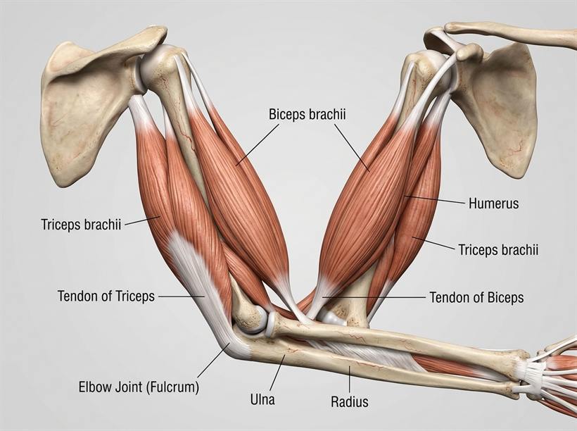
A. Bending (Flexing) the Arm at the Elbow
- The Biceps contracts (becomes short, thick, and bulges). The biceps acts as the flexor muscle.
- The Triceps relaxes (becomes long and thin).
- The contracted biceps pulls the radius and ulna upward toward the humerus, decreasing the elbow joint angle.
B. Straightening (Extending) the Arm at the Elbow
- The Triceps contracts (becomes short, thick, and firm). The triceps acts as the extensor muscle.
- The Biceps relaxes (becomes long and thin).
- The contracted triceps pulls the ulna downward, straightening the arm and increasing the elbow joint angle.
5. The Primary 7 Wooden Arm Teaching Model
The wooden arm model is an essential mechanical teaching tool constructed from local classroom materials to demonstrate antagonistic muscle action as a third-class lever:
| Model Component | Human Arm Part Represented | Function in the Demonstration |
|---|---|---|
| Upper piece of wood / plywood | Humerus (Upper arm bone) | Serves as the fixed anchor bone. |
| Lower piece of wood / plywood | Radius and Ulna (Forearm bones) | Serves as the movable lever arm. |
| String / Cord / Thread | Biceps Muscle (Flexor muscle) | Pulls and shortens to flex/bend the forearm upward. |
| Rubber band / Elastic strip | Triceps Muscle (Extensor muscle) | Stretches during flexion and contracts to straighten forearm. |
| Bolt / Screw / Hinge | Elbow Joint (Fulcrum / Hinge joint) | Serves as the pivot point for one-plane movement. |
| Drawing pins / Small nails | Tendons | Fasten and anchor the string/rubber band to the wood. |
When a question asks: "Name the two muscles found in the human arm", UNEB examiners strictly require the specific anatomical names: (i) Biceps and (ii) Triceps.
Reject: Writing "Flexor and Extensor" — flexor and extensor describe actions/roles, not the names of the muscles!
Posture, Bone Health, Diseases & Musculoskeletal Disorders
Theme: THE HUMAN BODY
Topic 1: THE MUSCULAR AND SKELETAL SYSTEM
Subtopic 5: Posture Ergonomics, Skeletal Diseases, Fractures & First Aid • Source: Primary 7 Science National Curriculum Textbook & P.7 Science Comprehensive Guide
1. Body Posture & Ergonomics
Posture is the position or carriage in which a person holds their body when sitting, standing, walking, sleeping, writing, or lifting objects.
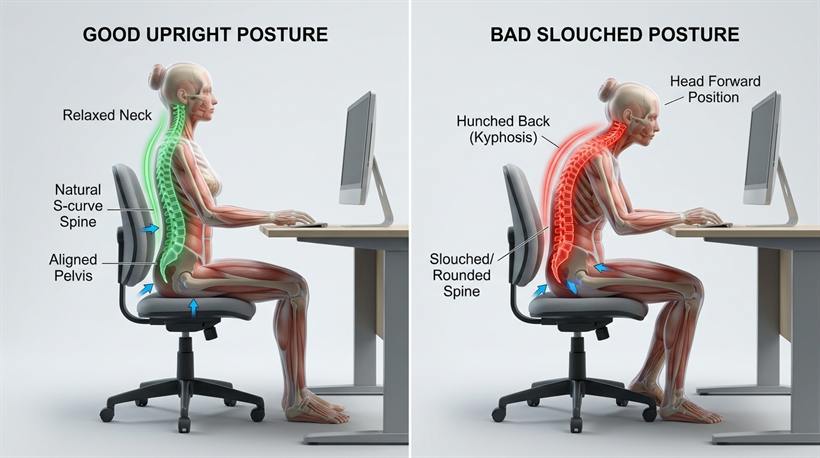
A. Rules for Maintaining Good Body Posture:
- Sitting at a Desk: Sit upright with the spine straight against the backrest of the chair; keep both feet flat on the floor; distribute body weight evenly on both buttocks; avoid slouching, hunching the shoulders, or bending the neck close to the desk.
- Standing: Stand tall with the head up, chin tucked in, shoulders relaxed and squared, chest held high, and weight balanced equally on both feet.
- Walking: Walk upright with head held high; look straight ahead; swing arms naturally and rhythmically; avoid stooping or shuffling the feet.
- Lifting Heavy Loads Correctly (CRITICAL):
✔ The Correct Method: Stand close to the heavy load; bend your hips and knees into a deep squat while keeping the back straight; grasp the load firmly; lift the load by straightening your legs and pushing upward with your strong thigh muscles (quadriceps).
❌ The Dangerous Method: Bending over at the waist with straight legs. This concentrates immense mechanical leverage and stress on the lumbar vertebrae, risking slipped intervertebral discs, severe nerve pinching, and lifelong backache.
B. Advantages of Maintaining Good Posture:
- Prevents bone deformities and abnormal spinal curvature (such as hunchback or scoliosis).
- Prevents chronic backache, neck strain, and chest pains.
- Allows the lungs to expand fully during breathing, ensuring maximum oxygen intake.
- Aids smooth digestion by preventing compression of abdominal organs (stomach and intestines).
- Enables easy, unconstricted blood circulation throughout the body.
- Gives an alert, dignified, smart, and confident appearance.
2. Diseases of the Skeletal and Muscular Systems
The human muscular and skeletal systems can be attacked by nutritional deficiencies, bacteria, and viruses:
| Disease | Cause & Transmission | Signs, Symptoms & Effects | Prevention & Control |
|---|---|---|---|
| 1. Poliomyelitis (Polio) | Poliovirus. Waterborne virus spread via drinking contaminated water, dirty food, or houseflies. |
Attacks motor nerves in spinal cord, causing paralysis of limbs, muscle wasting, and permanent lameness. | Immunization with Polio vaccine (OPV/IPV) at birth, 6, 10, 14 weeks; drinking boiled water; proper latrine hygiene. (Victims use crutches). |
| 2. Tuberculosis of the Spine / Bones (Spinal TB) | Mycobacterium tuberculosis. Airborne droplets or drinking unboiled milk from tubercular cows. |
Persistent severe backache, paralysis of legs, and formation of a hunchback (kyphosis) on the spine. | Immunization with BCG vaccine at birth on right upper arm; boiling/pasteurizing milk; early hospital antibiotic treatment. |
| 3. Rickets | Nutritional deficiency of Vitamin D, Calcium, or Phosphorus. | Soft, pliable bones; bow-legs (ox-bow legs); knock-knees; pigeon chest; delayed tooth eruption; swollen joints. | Morning sunbathing (sunlight UV stimulates skin to synthesize Vitamin D); diet rich in milk, eggs, small fish with bones (mukene), and cod liver oil. |
| 4. Tetanus (Lockjaw) | Clostridium tetani bacteria. Spores enter through dirty cuts, punctures from rusty nails, or dirty cord severance. |
Violent muscle spasms, stiff neck, arched back, and lockjaw (jaw muscles become rigid, preventing suckling or eating). | Immunization with DPT-HepB-Hib vaccine in infants and Tetanus Toxoid (TT) in expectant mothers; cleaning and dressing wounds immediately. |
| 5. Leprosy | Mycobacterium leprae. Spread through close body contact or via cockroaches. |
Pale skin patches devoid of feeling/sensation, nerve damage, and deformed/shortened toes and fingers. | Avoid close skin contact with untreated patients; maintain domestic hygiene; multi-drug therapy (MDT) at hospital. |
| 6. Osteoarthritis | Degenerative wear-and-tear of articular cartilage and loss of synovial fluid in aging joints. | Severe joint pain, swelling, stiffness, and bone rubbing friction, especially in the knees, hips, and fingers. | Regular gentle exercise, weight management to reduce pressure on joints, and medical pain relief. |
3. Bone Fractures & First Aid Management
A fracture is a broken or cracked bone in the body.
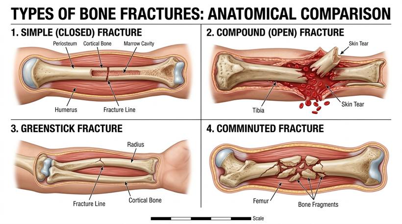
A. Major Types of Bone Fractures:
- Simple (Closed) Fracture:
The bone breaks or cracks inside the body, but the surrounding skin remains intact and unbroken. There is no external bleeding and low risk of bacterial infection.
- Compound (Open) Fracture:
The broken ends of the bone tear through muscle tissue and pierce outside through the skin. This creates an open bleeding wound with protruding bone, carrying a very high risk of bacterial infection and severe haemorrhage.
- Greenstick Fracture:
The bone bends and cracks on one side without breaking completely into two pieces (resembling a snapped green twig). This fracture is most common in young children because their bones contain more flexible cartilage and are soft and pliable.
- Comminuted Fracture:
The bone is shattered or crushed into three or more small pieces/splinters. Usually caused by heavy mechanical impacts such as motor vehicle accidents.
- Depressed Fracture:
A broken bone fragment is driven inward below its normal level. Common in skull injuries caused by blows from blunt objects.
- Impacted Fracture:
The broken jagged ends of the bone are driven forcibly into each other by violent compression.
B. First Aid for Bone Fractures:
- Splints:
Rigid pieces of wood, bamboo, or stiff cardboard placed along either side of the broken limb and tied firmly with bandages.
Function: Keeps the broken bone in one fixed position (immobilizes the bone), preventing bone ends from moving, cutting blood vessels, piercing nerves, or tearing muscles. - Triangular Arm Sling:
A triangular piece of cloth tied around the patient's neck.
Function: Supports, elevates, and immobilizes a fractured arm or collarbone. - Crutches:
Wooden or aluminium walking supports.
Function: Reduce the body weight placed on an injured, fractured, or healing leg while walking. - Stretcher:
Used to transport casualties who cannot walk without moving their spine, pelvis, or legs.
- Plaster of Paris (P.O.P):
Applied in hospitals as a hard casting material to hold the fractured bone rigid and securely aligned until new bone tissue (callus) knits together.
4. Joint and Muscle Injuries
- Dislocation: The displacement of a bone from its normal joint position or socket.
- Sprain: The violent overstretching or tearing of a ligament at a joint.
- Strain: The overstretching or tearing of a muscle or tendon.
- R — REST: Immediately cease all physical movement and rest the injured limb.
- I — ICE: Apply an ice pack or cold water compress for 15–20 minutes to constrict blood vessels, reduce internal bleeding, ease pain, and minimize swelling.
- C — COMPRESSION: Wrap a firm elastic crepe bandage around the injured joint to provide support and limit internal fluid accumulation (do not tie too tightly to cut off circulation).
- E — ELEVATION: Raise the injured limb above the level of the heart to reduce blood pressure and swelling at the injury site.
- ❌ Mistake: Defining fracture as "a broken bone".
✔ Examiner Correction: A fracture is "a broken or cracked bone in the body". - ❌ Mistake: Stating "Polio causes lameness" when asked about skeletal effects.
✔ Examiner Correction: State: "Polio causes paralysis of limbs". - ❌ Mistake: Writing "Tuberculosis" when asked for a bone disease.
✔ Examiner Correction: Must specify: "Tuberculosis of the spine / bones" (or Spinal TB). - Spelling Accuracy: Examiners immediately penalize misspelled keywords:
• Write Casualty (NOT Casuality)
• Write Tetanus (NOT Tetenus)
• Write Backbone as one word (NOT Back bone)
• Write Vertebral column (NOT Vertebral colum)
You have completed Topic 1!
Ready to test what you've learned? Challenge yourself with 8 authentic Primary Leaving Examination practice questions.